Diptera.info :: Identification queries :: Diptera (adults)
|
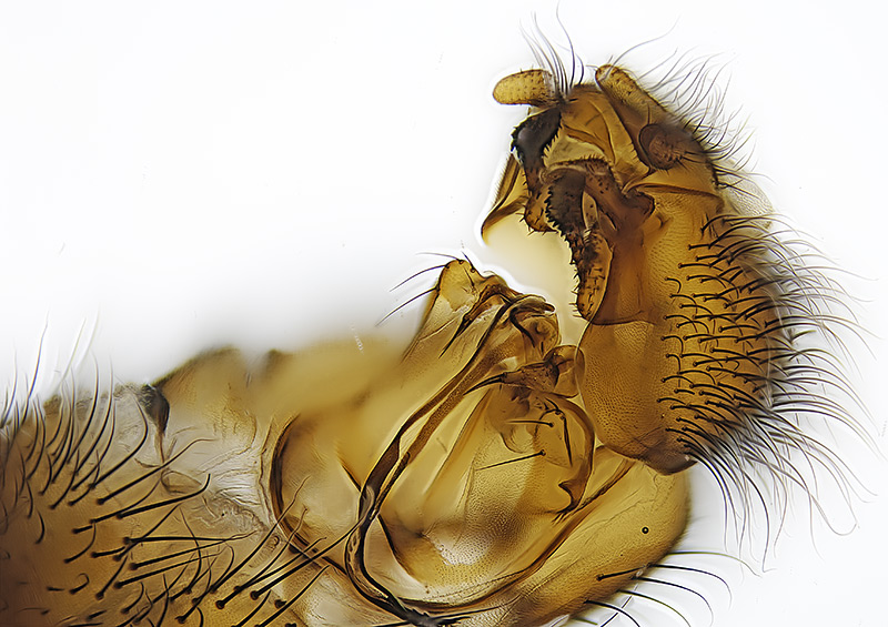
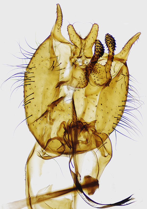
Heleomyzidae: Scoliocentra sackeni (Garrett) - male genitalia
|
|
| Tony T |
Posted on 18-04-2008 03:21
|
|
Member Location: New Brunswick, Canada Posts: 664 Joined: 08.02.07 |
From this male SEE HERE Andrzej: what a complex genitalia, hope the orientation is OK. Tony T attached the following image:  [171.18Kb] Edited by Tony T on 22-04-2008 10:55 |
|
|
|
| Susan R Walter |
Posted on 18-04-2008 13:15
|
|
Member Location: Touraine du Sud, central France Posts: 1802 Joined: 14.01.06 |
Phwoaw. This is one instance where a photograph is much better than a drawing.
Susan |
| phil withers |
Posted on 18-04-2008 13:45
|
|
Member Location: Lyon, France Posts: 521 Joined: 04.03.08 |
Excellent pic, but confusing. There appear to be two structures on the left side of the epandrium and only one on the right (maybe the orientation). I'm excluding the surstyli at the top; in any event this is not a Suillia pattern (and I thought the head shot wasn't Suillia either). You need to run this through Gill's key to N. American heleomzids (which I used to have but my ex-wife destroyed in a fit of...guess) to get closer to the genus. |
|
|
|
| Tony T |
Posted on 18-04-2008 14:52
|
|
Member Location: New Brunswick, Canada Posts: 664 Joined: 08.02.07 |
Thanks Susan & Phil. Andrzej wasn't too impressed, wants me to separate out all the little bits - moth dicks are so much easier  |
|
|
|
| Susan R Walter |
Posted on 18-04-2008 21:41
|
|
Member Location: Touraine du Sud, central France Posts: 1802 Joined: 14.01.06 |
Well, of course, Andrzej will have reason and is the expert for this family. I was really making a more general comment about good photographs versus drawings for certain features. For instance, based on my experience with male Bombus genitalia, I find using the photographs in Edwards and Jenner much easier than the drawings in Benton.
Susan |
| Tony T |
Posted on 19-04-2008 16:41
|
|
Member Location: New Brunswick, Canada Posts: 664 Joined: 08.02.07 |
phil withers wrote: There appear to be two structures on the left side of the epandrium and only one on the right (maybe the orientation). I'm excluding the surstyli at the top; in any event this is not a Suillia pattern (and I thought the head shot wasn't Suillia either).. Genital capsule flattened, ventral view. Does this help? Tony T attached the following image:  [170.55Kb] |
|
|
|
| Andrzej |
Posted on 20-04-2008 18:22
|
|
Member Location: Poland Posts: 2422 Joined: 05.01.06 |
Hmm, I am in trouble  I have determined the female as Heleomyza. cf. difficilis but the genitalia of the previous determined male look like Scoliocentra sensu stricto. Tony could you check by the male specimen the number of prosternal bristles (1 pair or more than 2 pairs) and the hind femur (if without brush-like spines or with such bristles on the lower inner side) ??. Regards, Andrzej |
|
|
|
| Nosferatumyia |
Posted on 20-04-2008 22:20
|
|
Member Location: Posts: 3550 Joined: 28.12.07 |
Tony, I am impressed again. No microscope used? Have you explained your techics in this case already? Just a reverted wideangle objective? And the system of sledges to move the camera slowly you have depicted? I think NO BRAND NEW ready camera+microscope system could achieve such a quality... Damned tasty... 
Val |
|
|
|
| Tony T |
Posted on 21-04-2008 00:53
|
|
Member Location: New Brunswick, Canada Posts: 664 Joined: 08.02.07 |
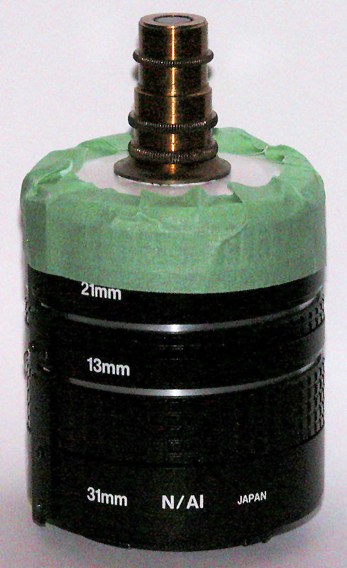
Andrzej wrote: Tony could you check by the male specimen the number of prosternal bristles (1 pair or more than 2 pairs) and the hind femur (if without brush-like spines or with such bristles on the lower inner side) ??. Regards, Andrzej Very cleary there is just 1 pair of prosternal bristles. Unfortunately the hind legs broke off when I removed the abdomen. I have the legs but not sure which is outer or inner. Either way, I cannot see any spines on the lower part of the femur. Valery wrote "No microscope used? Just a reverted wideangle objective? And the system of sledges to move the camera slowly you have depicted?" No microscope - not necessary! But for this image I used a 10x microscope objective connected to some extensions tubes (see photo) and then connected to a bellows, the bellows attached to the camera. The total length of extension was 160mm which I believe is the length that these microscope objectives are designed for. For such small specimens, in this case the genital capsule if just greater than 1mm, one has to use a microscope objective. For larger specimens a reversed wide angle lens is OK and for an entire fly a regular lens on extensions tubes or bellows. One can move either the camera or the specimen. I always move the specimen. None of my techniques are my originals, they are all standard methodology for macrophotography. Tony T attached the following image:  [84.35Kb] |
|
|
|
| Nosferatumyia |
Posted on 21-04-2008 06:02
|
|
Member Location: Posts: 3550 Joined: 28.12.07 |
Tony, thanx a lot - I am totally a newbie  in that sort of things, I used to draw all that stuff in ink until 2005. I cannot say that the microphotography is less time consuming, but the results are so miraculous... in that sort of things, I used to draw all that stuff in ink until 2005. I cannot say that the microphotography is less time consuming, but the results are so miraculous...
Val |
|
|
|
| Andrzej |
Posted on 22-04-2008 10:08
|
|
Member Location: Poland Posts: 2422 Joined: 05.01.06 |
So, it is clear !. It is Scoliocentra sackeni (Garrett  Andrzej |
|
|
|
| Tony T |
Posted on 22-04-2008 10:53
|
|
Member Location: New Brunswick, Canada Posts: 664 Joined: 08.02.07 |
Thanks Andrzej. I must check and see if I have any other males |
|
|
|
| Jump to Forum: |


